
Understanding Posterior Tibial Tendonitis
Understanding this condition involves recognizing its progressive nature; early intervention, utilizing a structured exercise program, is crucial to prevent irreversible foot structure changes.
PTTD management often centers around a protocol combining orthotics and targeted exercises, specifically designed to strengthen the posterior tibialis muscle.
A consistent Home Exercise Program (HEP), completed 5-7 times weekly, is vital throughout rehabilitation, emphasizing posterior tibialis focused movements.
What is the Posterior Tibial Tendon?
The posterior tibial tendon is a crucial structure, acting as a dynamic support for the foot’s arch. Originating from the calf muscle, it travels down the inner leg and attaches to bones within the foot.
Its primary responsibility is to maintain the arch’s integrity during weight-bearing activities like walking and jumping. A healthy tendon effectively supports the foot, enabling smooth and stable movement.
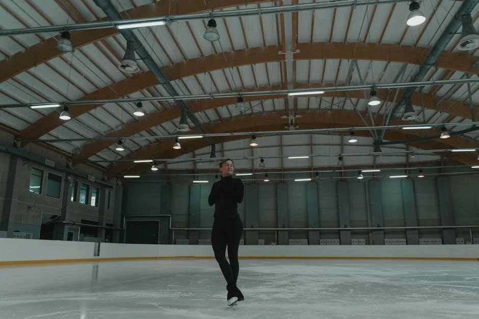
However, this tendon is susceptible to injury, leading to Posterior Tibial Tendon Dysfunction (PTTD). Understanding its function is key to appreciating the impact of dysfunction and the importance of targeted exercise and rehabilitation. Strengthening this tendon is vital for foot stability.
Causes of Posterior Tibial Tendonitis
Posterior tibial tendonitis, or PTTD, develops from a combination of factors, often involving gradual degeneration rather than a single acute injury. Overuse, particularly in activities with repetitive impact, is a significant contributor.
Risk factors include high-impact exercise, obesity, and structural foot abnormalities like flat feet. An acute injury, such as a fall, can also initiate the process.
This degeneration weakens the tendon, hindering its ability to support the arch. Early intervention, including a tailored exercise program, is crucial to slow progression and prevent irreversible structural changes. Addressing biomechanical issues is also key.
Symptoms of Posterior Tibial Tendonitis
Symptoms typically begin gradually, with pain and swelling along the inner side of the ankle and foot. Individuals may notice a progressive flattening of the arch, leading to a change in gait.
Pain often worsens with activity and improves with rest, initially. As the condition progresses, pain can become constant, and the foot may become rigid.
Early diagnosis and a structured exercise program are vital to manage symptoms. The ability to perform single-leg jumps without pain indicates a positive response to rehabilitation, but professional assessment is crucial.

Diagnosis and Assessment
Diagnosis relies on a thorough physical examination and may include imaging tests like X-rays and MRI to evaluate tendon integrity and foot structure.
Physical Examination Techniques
Assessment begins with a detailed patient history, focusing on activity levels and symptom onset. Palpation along the posterior tibial tendon identifies tenderness. The Single Limb Stance Test assesses arch collapse; a noticeable flattening suggests dysfunction.
Foot and ankle range of motion are evaluated, noting any limitations. A “too many toes” sign – observing excessive outward tilting of the foot – is indicative. Neurological and vascular assessments rule out other contributing factors. Careful observation of gait reveals compensatory patterns.
Strength testing focuses on calf muscles and intrinsic foot muscles. These techniques collectively help determine the severity and stage of PTTD.
Imaging Tests (X-rays, MRI)
X-rays are initially used to rule out other conditions and assess for arthritic changes. However, they often appear normal in early PTTD. As the condition progresses, X-rays may reveal flattening of the arch and bone spurs.
MRI provides a more detailed view of the posterior tibial tendon itself, visualizing tears, inflammation, and tenosynovitis. It’s crucial for confirming the diagnosis and assessing the extent of tendon damage.
MRI also helps evaluate surrounding structures, like ligaments and cartilage. This imaging allows for accurate staging of PTTD, guiding treatment decisions and monitoring progress.

Non-Surgical Treatment Options
Non-surgical approaches prioritize rest, activity modification, and targeted exercises to strengthen the posterior tibialis, often combined with orthotic support for arch control.
Rest and Activity Modification
Initial management of posterior tibial tendonitis necessitates a period of rest, crucially avoiding activities that exacerbate pain, such as prolonged standing, walking, or high-impact exercises. Activity modification is paramount; transitioning to lower-impact alternatives like swimming or cycling can maintain fitness without stressing the tendon.
Gradually decreasing activity levels allows inflammation to subside, paving the way for a structured rehabilitation program. Complete rest isn’t always ideal, as prolonged immobilization can lead to muscle weakness and stiffness, hindering recovery. Therefore, a balanced approach—reducing aggravating activities while maintaining gentle movement—is recommended.
Listen to your body and adjust activity levels accordingly, prioritizing pain-free movement as a guiding principle.
Ice and Compression
Applying ice to the affected area is a cornerstone of early posterior tibial tendonitis management, effectively reducing inflammation and alleviating pain. Ice packs should be applied for 15-20 minutes at a time, several times daily, especially after activity. Always use a cloth barrier to protect the skin.
Compression, utilizing an elastic bandage, further aids in minimizing swelling and providing support to the injured tendon. Ensure the bandage isn’t too tight, as this can impede circulation. Combining ice and compression offers synergistic benefits, accelerating the healing process.
These conservative measures are often implemented alongside rest and activity modification.
Orthotics and Arch Support
Orthotics play a crucial role in managing posterior tibial tendonitis by providing essential arch support and correcting biomechanical imbalances. Custom or over-the-counter arch supports help redistribute pressure, reducing stress on the inflamed tendon.
These supports limit excessive pronation, a common contributing factor to the condition. Proper footwear, combined with orthotics, creates a stable environment for the foot during activity.
Consistent use, even during low-impact activities, is recommended. Orthotics are often integrated into a comprehensive treatment plan alongside exercise and other conservative measures.
Posterior Tibial Tendonitis Exercises, Phase 1 (Early Stage)
Phase 1 focuses on gentle movements: towel curls, double leg calf raises, and alphabet tracing with the foot, initiating a pain-free recovery.
Towel Curls
Towel curls are a foundational exercise in Phase 1, designed to gently activate the posterior tibialis muscle without placing excessive stress on the injured tendon. To perform this exercise, sit comfortably with your foot flat on a towel placed on the floor.
Using only your toes, curl the towel towards you, drawing it closer with a controlled motion. Focus on engaging the muscles on the inner side of your ankle and foot. Slowly release and repeat the curling motion 10-15 times.
This exercise helps improve muscle strength and coordination, preparing the tendon for more advanced rehabilitation exercises. Ensure the movement is pain-free; stop if you experience any discomfort.
Calf Raises (Double Leg)
Double leg calf raises are a crucial early-stage exercise, strengthening the calf muscles which support the posterior tibialis tendon. Stand with your feet flat on the floor, shoulder-width apart, and maintain a slight bend in your knees.
Slowly rise up onto your toes, lifting your heels off the ground as high as comfortably possible. Focus on controlled movement, avoiding any jerky motions. Hold the raised position for a second, then slowly lower your heels back to the floor.
Repeat this exercise 10-15 times. This builds strength and endurance, preparing the lower leg for more challenging exercises in later phases of rehabilitation.
Alphabet Tracing with Foot
Alphabet tracing is a gentle, yet effective, exercise to improve ankle range of motion and posterior tibialis control. Sit comfortably with your leg extended, and slowly “write” each letter of the alphabet using your big toe as a pencil.
Focus on maintaining control throughout the movement, and avoid compensating with your entire leg. This exercise promotes proprioception – your body’s awareness of its position in space – and helps restore normal foot mechanics.
Perform one full alphabet cycle, repeating 2-3 times. This exercise is excellent for early-stage rehabilitation, enhancing coordination and preparing for weight-bearing activities.
Posterior Tibial Tendonitis Exercises ― Phase 2 (Intermediate Stage)
Phase 2 builds strength and endurance with exercises like single leg calf raises, heel walks, and toe walks, progressing from initial stabilization efforts.
Single Leg Calf Raises
Single leg calf raises are a crucial progression in posterior tibial tendonitis rehabilitation, building strength and endurance in the calf muscles and supporting tendon. Begin by standing on the affected leg, using a chair or wall for balance if needed.
Slowly rise up onto your toes, focusing on controlled movement and full range of motion. Hold briefly at the top, then slowly lower back down. Perform 3 sets of 10-15 repetitions, gradually increasing the repetitions as strength improves.
Ensure proper form – avoid rolling your ankle inward or outward. This exercise directly targets the muscles supporting the arch, aiding in restoring function and preventing further strain on the posterior tibialis tendon.
Heel Walks
Heel walks are an excellent intermediate-stage exercise for posterior tibial tendonitis rehabilitation, strengthening the anterior tibialis muscle and improving ankle dorsiflexion. To perform, lift your toes off the ground and walk forward solely on your heels for a designated distance – approximately 20-30 feet.
Maintain a controlled pace and focus on keeping your ankle stable. Perform 3 sets of heel walks, resting between each set. This exercise helps counteract the strain on the posterior tibialis by strengthening opposing muscle groups.
Pay attention to any pain; modify the distance or discontinue if discomfort arises.
Toe Walks
Toe walks represent another valuable intermediate-stage exercise within a posterior tibial tendonitis rehabilitation program, focusing on strengthening the calf muscles and promoting plantarflexion. To execute, lift your heels off the ground and walk forward exclusively on your toes for a distance of 20-30 feet.
Maintain a steady, controlled gait, concentrating on engaging the calf muscles throughout the movement. Complete 3 sets of toe walks, incorporating rest periods between each set. This exercise indirectly supports the posterior tibialis by enhancing overall ankle stability.
Monitor for any pain signals and adjust accordingly.
Posterior Tibial Tendonitis Exercises ― Phase 3 (Advanced Stage)
Phase 3 focuses on advanced strengthening and proprioception, incorporating eccentric heel drops, single-leg stance work, and targeted training for optimal foot stability.
Eccentric Heel Drops
Eccentric heel drops are a cornerstone of Phase 3 rehabilitation, specifically targeting the posterior tibialis tendon. Perform these by standing on a slightly elevated surface, like a step. Rise up onto your toes using both legs, then slowly lower the affected heel below the step’s level.
Control the descent, resisting the pull of gravity – this is the eccentric portion. Focus on a slow, controlled movement over 3-5 seconds. Begin with 3 sets of 10-15 repetitions, gradually increasing the number of sets and reps as tolerated. Ensure proper form to avoid re-injury; pain should be minimal and controlled.
This exercise strengthens the tendon during lengthening, promoting improved function and reducing pain.
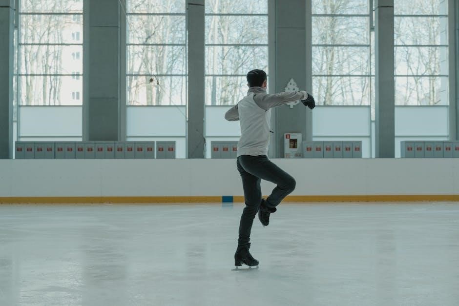
Balance Exercises (Single Leg Stance)
Single leg stance is a crucial component of Phase 3, enhancing proprioception and strengthening supporting muscles around the ankle and foot. Begin by standing on the affected leg, maintaining a slight bend in the knee. Hold this position for 30-60 seconds, aiming for stability without wobbling.
Progress by performing the exercise with your eyes closed, or on an unstable surface like a foam pad. Complete 3-5 repetitions per leg. Focus on engaging your core and maintaining proper alignment.
This exercise improves dynamic stability and prepares the foot for higher-level activities.
Proprioceptive Training
Proprioceptive training, vital in Phase 3, focuses on restoring the foot’s ability to sense its position in space. Begin with simple exercises like weight shifting, gently rocking forward, backward, and side-to-side while maintaining balance.
Progress to more challenging activities, such as standing on an unstable surface (foam pad or wobble board) and performing small, controlled movements. Incorporate reaching tasks while balancing to further challenge stability.
These exercises enhance neuromuscular control, improving coordination and reducing the risk of re-injury. Aim for 3-5 repetitions, focusing on controlled movements and maintaining balance.

Rehabilitation Protocol & Guidelines
Rehabilitation follows a phased approach, emphasizing consistent HEP adherence (5-7 times weekly) and progressive exercise loading, guided by pain and functional improvements.
Guidelines prioritize posterior tibialis strengthening and restoring foot/ankle stability, crucial for returning to high-level activity or sports.
Frequency and Duration of Exercises
Consistent adherence to the prescribed Home Exercise Program (HEP) is paramount for successful rehabilitation. Generally, exercises should be performed 5-7 times per week, allowing for adequate rest and recovery between sessions.
Initially, each exercise may be completed for 2-3 sets of 10-15 repetitions. As strength and pain levels improve, the number of sets and repetitions can be gradually increased. It’s crucial to listen to your body and avoid pushing through significant pain.
Duration of each exercise session will vary depending on the phase of rehabilitation, but typically ranges from 20-30 minutes. Regular review and re-emphasis of the HEP by a therapist ensures proper form and progression.
Progression Criteria
Advancement through rehabilitation phases hinges on meeting specific criteria, not simply time elapsed. A key indicator is the ability to perform exercises with minimal pain – ideally, no more than a 2/10 on a pain scale.
Successful completion of current exercise sets and repetitions with good form is essential. Functional improvements, such as improved balance (single leg stance) and walking mechanics, also signal readiness for progression.
The ability to jump on both legs without pain, and subsequently on the injured leg, demonstrates sufficient strength and stability. A therapist’s evaluation confirms these milestones before advancing to more challenging exercises.
Surgical Intervention
Surgery is considered when non-surgical methods fail, often involving tendon repair or transfer alongside calcaneal osteotomy to restore foot alignment.
Rehabilitation guidelines are crucial post-op, with a phased approach beginning immediately after the posterior tibial tendon repair.
When is Surgery Considered?
Surgical intervention for posterior tibial tendonitis isn’t the first line of defense; it’s typically reserved for cases where conservative treatments – rest, orthotics, and a dedicated exercise program – have proven ineffective over an extended period.
Specifically, surgery becomes a viable option when significant structural damage has occurred, leading to substantial foot deformity and persistent pain that severely limits daily activities.
If the tendon is severely torn or the foot has progressed to a rigid deformity, surgical reconstruction, like tendon repair or transfer with a calcaneal osteotomy, may be necessary to restore function and alleviate symptoms. The decision is made collaboratively between the patient and surgeon.
Surgical Procedures (Tendon Repair, Transfer)
Surgical options for posterior tibial tendon dysfunction aim to restore foot stability and alleviate pain. Tendon repair involves directly reattaching the torn tendon to its original insertion point on the navicular bone, if sufficient tissue quality exists.
However, often the tendon is too damaged for direct repair, necessitating a tendon transfer. This involves relocating another tendon, typically the flexor digitorum longus, to take over the function of the damaged posterior tibialis.
Frequently, a calcaneal osteotomy is performed concurrently to realign the heel bone and improve foot mechanics, optimizing the transferred tendon’s effectiveness and long-term outcome.

Post-Operative Rehabilitation
Post-operative care follows a phased approach, beginning with immobilization and gentle range-of-motion exercises, progressing to weight-bearing and strengthening as healing advances.
Rehabilitation guidelines emphasize gradual loading and adherence to a structured exercise plan, tailored to each phase of recovery post-repair or transfer.
Phase 1 (0-6 Weeks Post-Op)
Phase 1 focuses on protecting the surgical repair and minimizing swelling. Rehabilitation begins with strict non-weight bearing, utilizing crutches or a scooter, and immobilization in a cast or boot. Gentle range-of-motion exercises, focusing on ankle pumps and toe wiggles, are initiated to maintain circulation and prevent stiffness.
Exercises are performed within a pain-free range. Isometric exercises, contracting muscles without movement, may be introduced towards the end of this phase to begin activating the posterior tibialis without stressing the repair. Emphasis is placed on edema control with elevation and ice. The goal is to achieve a pain-free, protected healing environment, preparing for the next phase of rehabilitation.
Phase 2 (6-12 Weeks Post-Op)
Phase 2 transitions towards partial weight-bearing, as tolerated, often with continued use of a boot or orthotic. Exercises progress to include gentle stretching and strengthening of the calf muscles and intrinsic foot muscles. Towel curls and calf raises, initially double-leg, are introduced to begin restoring strength in the posterior tibialis.
Proprioceptive exercises, like weight shifting, are added to improve balance and coordination. Pain and swelling continue to be monitored closely, guiding progression. The focus remains on gradually increasing weight-bearing tolerance and restoring a more normal gait pattern, preparing for advanced strengthening in Phase 3.
Preventing Recurrence
Maintaining strength through ongoing exercises, wearing supportive footwear, and adhering to a consistent rehabilitation plan are key to preventing PTTD return.
Prioritize flexibility and strength, alongside appropriate orthotic support, for long-term foot and ankle stability.
Proper Footwear
Selecting appropriate footwear is paramount in both preventing and managing posterior tibial tendonitis. Shoes should offer substantial arch support and excellent motion control to minimize stress on the affected tendon. Avoid flat shoes, high heels, and worn-out footwear, as these exacerbate the condition.
Look for shoes with a firm heel counter to stabilize the foot and prevent excessive pronation. Consider consulting a podiatrist or specialist running store for personalized recommendations based on your foot type and activity level. Properly fitted shoes, combined with orthotics when needed, create a supportive foundation for effective rehabilitation and long-term tendon health.
Ongoing Exercise Program
Maintaining a consistent exercise program post-rehabilitation is crucial for preventing recurrence of posterior tibial tendonitis. Continue performing the prescribed exercises – towel curls, calf raises, and balance work – 5-7 times per week, even after symptom resolution.
Focus on strengthening the posterior tibialis muscle and improving proprioception. Gradually increase exercise intensity and duration as tolerated. Incorporate activities that challenge balance and stability. Regular exercise, coupled with proper footwear and activity modification, ensures long-term tendon health and minimizes the risk of future complications.